Why Picosecond Laser Outperforms Traditional Nanosecond Laser: A Deep Tech Clinical Protocol
Clinic Pain Points: The Limitations of Nanosecond Lasers in Modern Aesthetics
For over a decade, Q-switched nanosecond lasers have been the clinical standard for tattoo removal and pigmentary disorders. However, clinics increasingly face a ceiling: stubborn multi-colored tattoos, paradoxical hyperpigmentation in Fitzpatrick Skin Types IV-VI, and lengthy treatment protocols. The physical mechanism of selective photothermolysis using nanosecond pulses relies primarily on photothermal expansion to shatter targets, generating significant heat that risks collateral thermal damage, post-inflammatory hyperpigmentation (PIH), and scarring. This limits energy fluence (J/cm²) and requires multiple sessions. As patient expectations shift toward faster clearance with minimal downtime, the picosecond laser has emerged as a transformative clinical alternative, leveraging peak power in the trillionths of a second to induce a photoacoustic — rather than primarily thermal — effect.
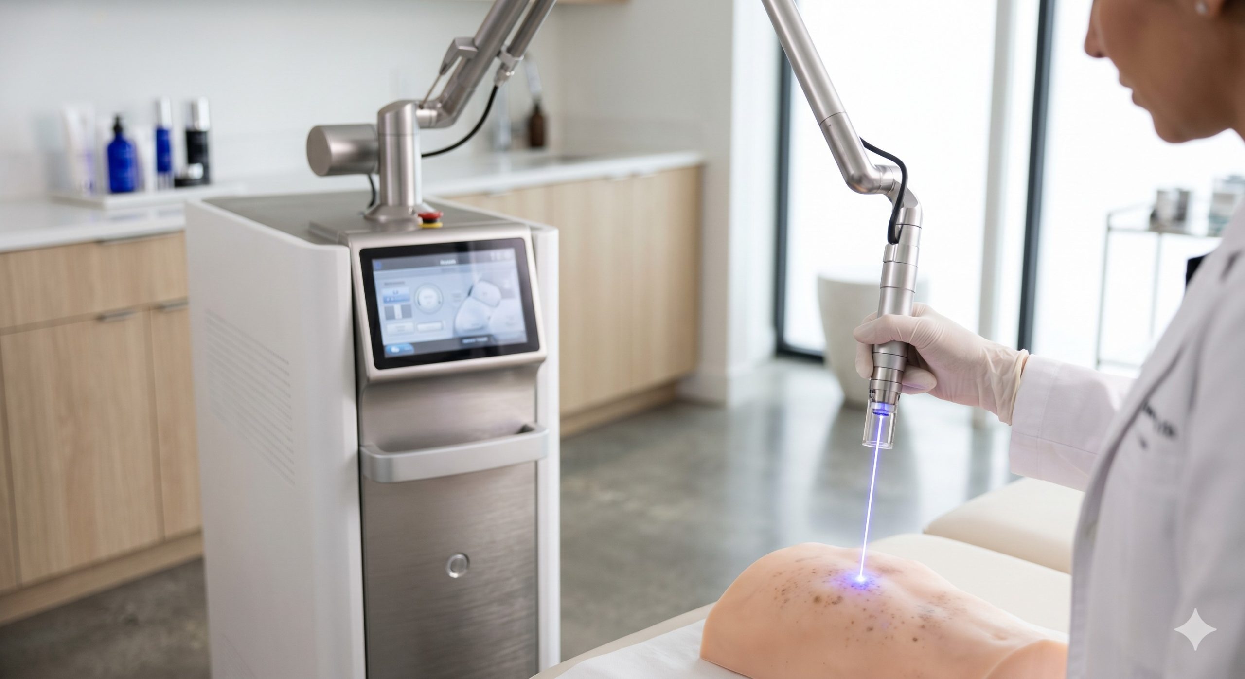
Physical Mechanism Deep Dive: Photoacoustic vs. Photothermal Interaction
The fundamental distinction lies in pulse width. A picosecond laser delivers energy in 350–750 picoseconds (10⁻¹² seconds), whereas traditional nanosecond lasers operate at 5–100 nanoseconds (10⁻⁹ seconds). This 10–100x shorter pulse duration generates immense peak power (gigawatts vs. megawatts), creating a laser-induced optical breakdown (LIOB) confined within the target chromophore — melanosomes or tattoo ink particles. The rapid thermoelastic expansion produces a plasma shockwave that mechanically fragments targets into dust particles (< 1 micron), enabling phagocytic clearance within weeks. Conversely, nanosecond pulses generate slower thermal expansion, often leaving larger residual particle fragments (5–10 microns) and requiring higher fluence to achieve similar clearance, thereby increasing the risk of epidermal injury.
Critical Output Metrics for Clinical Evaluation
When comparing devices, clinic decision-makers must analyze three interdependent parameters: pulse width (ps), energy density/fluence (mJ/cm² or J/cm²), and spot size (mm). A shorter pulse width allows lower fluence to achieve the same threshold fluence (Fth), preserving the epidermis. For example, nanosecond systems often require 2–5 J/cm² for black ink, while picosecond systems achieve equivalent clearance at 0.5–1.5 J/cm². However, not all picosecond labels are equal: true picosecond devices maintain a pulse width under 1,000 ps; hybrid systems that blend nanosecond pedestals significantly reduce the photoacoustic advantage. Clinical evidence also confirms that wavelengths — 532nm (superficial red/orange), 755nm (green/blue/ melanin), and 1064nm (deep black/dark tattoos and melasma) — must be paired with appropriate spot sizes (2–8mm) to match target depth and lesion geometry.
| Key Parameter | Picosecond Laser (True Ps) | Traditional Nanosecond Laser (Q-Switched) |
|---|---|---|
| Pulse Width (Typical) | 350 – 750 picoseconds | 5 – 100 nanoseconds |
| Peak Power (Typical) | 1.5 – 2.5 GW | 50 – 300 MW |
| Primary Mechanism | Photoacoustic / Shockwave | Photothermal / Thermal Expansion |
| Fluence for Black Ink | 0.5 – 1.5 J/cm² | 2.0 – 5.0 J/cm² |
| Particle Size After Lysis | < 1 micron (dust-like) | 5 – 10 microns (fragments) |
| Epidermal Thermal Damage Risk | Low (superior for Skin Types IV-VI) | Moderate to High (PIH risk) |
| Typical Spot Sizes | 2mm – 8mm (interchangeable) | 1.5mm – 6mm |
| FDA Indications | Tattoo, Pigmented lesions, Acne scars (fractional) | Tattoo, Pigmented lesions |
| Handpiece Lifespan (Shots) | 300,000 – 500,000 | 100,000 – 200,000 |
Epidermal Protection & Cooling Systems: Preventing PIH in Darker Skin
For clinics treating high volumes of Fitzpatrick Skin Types IV-VI, the superior safety profile of picosecond lasers is a compelling differentiator. The reduced thermal relaxation time minimizes residual heat diffusion into basal keratinocytes, substantially lowering the risk of post-inflammatory hyperpigmentation (PIH). To further safeguard the epidermis, high-end picosecond platforms integrate advanced cooling mechanisms:
- Sapphire contact cooling (typically -5°C to +5°C) with real-time temperature monitoring via thermistors.
- Cryogen spray cooling (CSC) — precisely timed 30–80ms spurt prior to laser pulse for superficial lesions.
- Imported dual-cycle water chillers (maintaining ±1°C stability) for high-throughput workflows.
These systems maintain epidermal temperatures below 40°C during treatment, allowing clinicians to safely increase fluence by 20–40% over nanosecond protocols, accelerating clearance without blistering. ISO 13485-certified manufacturers provide validation data on cooling uniformity across the entire spot area — a critical but often overlooked specification.
Clinical Indications & Fitzpatrick-Specific Protocols
The versatility of picosecond technology extends beyond tattoos. Leading Medical CE and FDA-cleared platforms now offer indications for:
- Benign pigmented lesions: Lentigines, ephelides, café-au-lait macules, and Hori’s nevus.
- Acne scars & textural improvement: The laser-induced collagen remodeling (LICR) effect using a diffractive lens array (DLA) or fractal lens — a lower-fluence, large-spot (6–10mm) mode that creates microscopic treatment zones in the dermis without ablating the epidermis.
- Melasma: Fractional picosecond modes with 1064nm and low fluence (0.3–0.7 J/cm²) have shown significant improvement in mixed-type melasma with minimal rebound hyperpigmentation compared to nanosecond or IPL therapies.
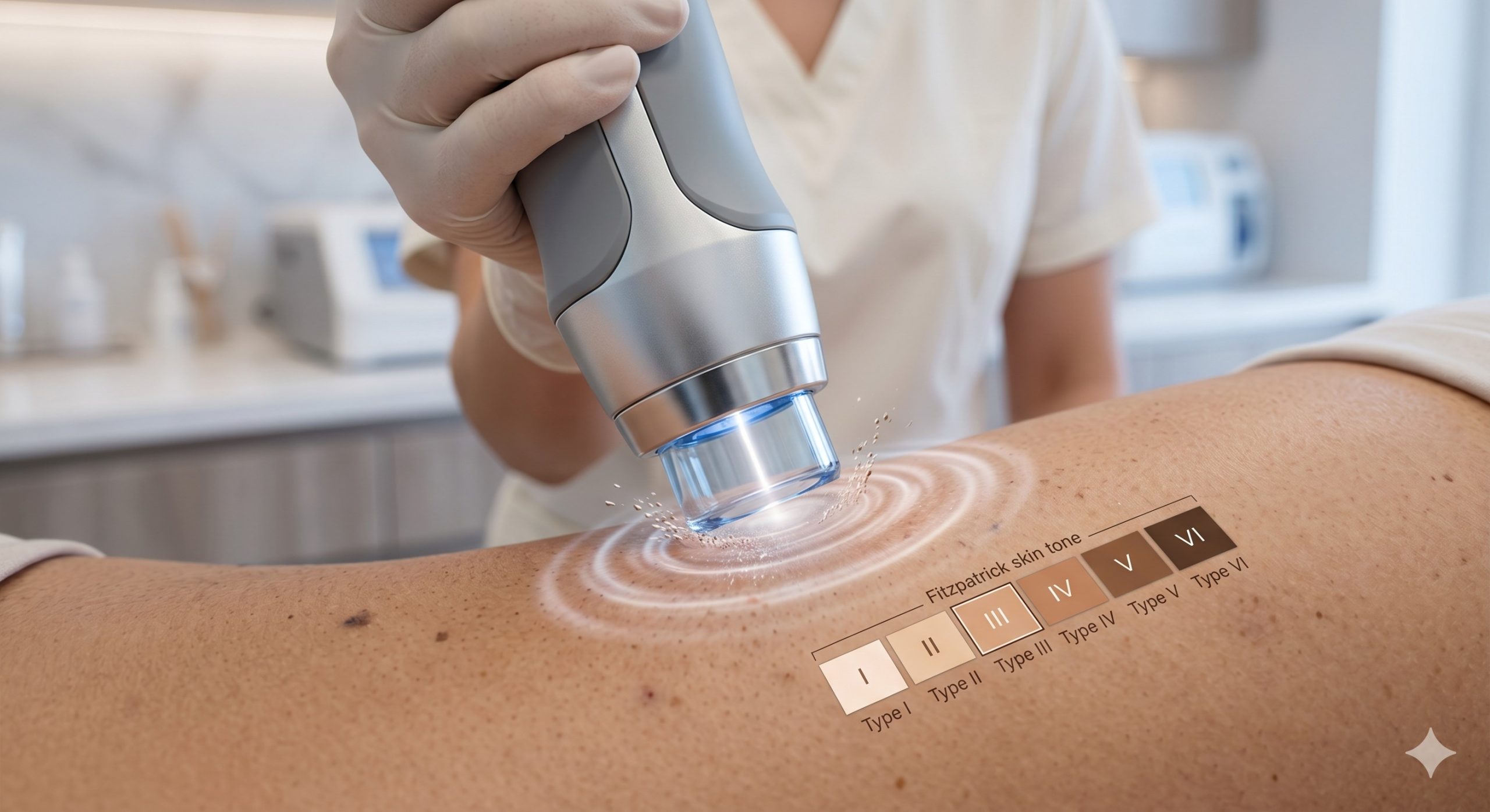
For Fitzpatrick Skin Types I-III, clinicians can utilize higher fluences and shorter wavelengths (532nm) aggressively. For Types IV-VI, start with 1064nm, 6mm spot, fluence 0.7–1.2 J/cm², and perform a test spot at 48 hours. Always use topographic anesthesia (not injection, which distorts tissue optics) for patient comfort.
Clinic ROI Analysis: From CapEx to Lifetime Profitability
While the upfront capital expenditure (CapEx) for a medical-grade picosecond laser (typically $60,000–$120,000 USD) is higher than a nanosecond system ($25,000–$50,000), the return on investment (ROI) analysis shifts in the picosecond’s favor within 12–18 months in a high-throughput clinic. Key drivers include:
- Higher revenue per session: Patients pay a 30–50% premium for ‘faster clearance with less risk’ — average picosecond tattoo removal session: $400–$800 vs. $250–$450 for nanosecond.
- Reduced consumables & TCO: True picosecond handpieces with imported laser bars (e.g., from JDSU or Coherent) deliver 300,000–500,000 shots before degradation, compared to 100,000–200,000 shots for nanosecond OEM modules. No flashlamps or dye cuvettes to replace quarterly.
- Treatment speed: Large spot sizes (up to 8mm) and repetition rates up to 10Hz allow full back tattoo coverage in 15 minutes vs. 40 minutes with a 3–4mm nanosecond spot — tripling daily patient throughput.
- Lower Malpractice risk & patient satisfaction: Reduced PIH and scarring rates lead to better reviews, more referrals, and fewer complication management costs.
Clinics should verify CE MD Class IIb or III certification, FDA 510(k) clearance for specific indications, and request independent clinical data from the manufacturer. A device without published human studies on at least 50 subjects is a liability, not an asset.
Executive Verdict: Strategic Adoption for Forward-Thinking Clinics
The transition from nanosecond to picosecond laser technology is not a marginal improvement — it represents a paradigm shift in laser-tissue interaction. For clinics seeking to dominate the tattoo removal, pigmentation, and scar revision market, investing in a dual-wavelength (755nm & 1064nm) or triple-wavelength picosecond platform with integrated cooling and fractional handpieces offers the broadest clinical utility and highest patient retention. However, technology alone does not guarantee outcomes. Invest heavily in clinical training, develop standardized photo-documentation protocols, and market the ‘no downtime’ advantage. As AI-driven treatment planning and real-time skin response monitoring enter the market, only systems with open-architecture software and validated picosecond engines will remain relevant. The question is no longer ‘if’ but ‘when’ your clinic will upgrade.